Discover three game-changing advancements that are reshaping how Albertans experience their cancer journeys.
By Cailynn Klingbeil
Seeing cancer differently
Dr. Kristi Baker is using a new instrument to examine patient samples, aiming to better understand and treat advanced colon and lung cancers.
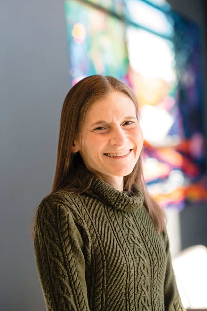
Dr. Kristi Baker is an associate professor in the departments of oncology and medical microbiology and immunology at the University of Alberta. She describes the new instrument her team uses inside a room at the university, known as the Cell Imaging Core, as “a really fancy microscope” — a purple machine paired with a box that circulates fluids through slides of tissue samples. Where a standard microscope might let a researcher examine one or two markers in a tissue sample at a time, this advanced multiplex imaging platform — known as the Akoya Phenocycler Fusion 2.0 — can examine almost 100 markers at once.
That instrument’s advanced capability is helping Baker’s team take an unprecedented look at immune cells inside colon and lung tumours. Using one of only a few advanced multiplex imaging platforms in Canada, they want to understand why these cancers spread and why some patients respond to treatment while others don’t — insights that could eventually inform new diagnostic tools and treatments.
Supported by the Alberta Cancer Foundation’s Game Changer competition, Baker’s research focuses on advanced-stage colon and lung cancers, two cancers with a low survival rate. While primary tumours can often be treated, it is the spread of cancer cells, known as metastasis, that is responsible for most cancer-related deaths.
“We really want to be able to compare between the primary tumour and the metastasis and [ask], do we see similar types of immune cells there, and if so, are they doing similar types of things?” says Baker.
Inside immune cells
The lungs and intestines are unlike most other organs in the body, Baker explains. They’re constantly exposed to outside substances, from the air we breathe to the food we eat, so the immune cells that live there have to be more tolerant.
They can’t react to every foreign particle they encounter, or the body would constantly be fighting itself. But that natural tolerance may also make it easier for cancer to hide from immune system defences. Baker hopes understanding these immune cells will reveal why advanced colon and lung cancers are often so hard to treat, particularly with immunotherapy.
The new multiplex imaging platform makes that investigation possible.
“We can actually look at different types of immune cells and different types of molecules and receptors that are present in the tumour. And we can see where they are in relation to other cells,” Baker says.
The work is a team effort, Baker adds, with graduate and undergraduate students doing much of the day-to-day lab work. It also depends on patients who donate leftover tissue after surgery, knowing that donation may not benefit them directly but could improve care for future patients.
The research project is still in its discovery phase. Since receiving the imaging instrument in January 2025, the team has optimized protocols and prepared samples. Next comes large-scale analysis with computational researchers.
Ultimately, Baker hopes her work helps identify which patients will respond to which treatments and find new ways of activating immune cells that currently fail to recognize advanced cancers.
A preventative approach
Dr. Claire Temple-Oberle’s work reduces one of cancer surgery’s common side effects.
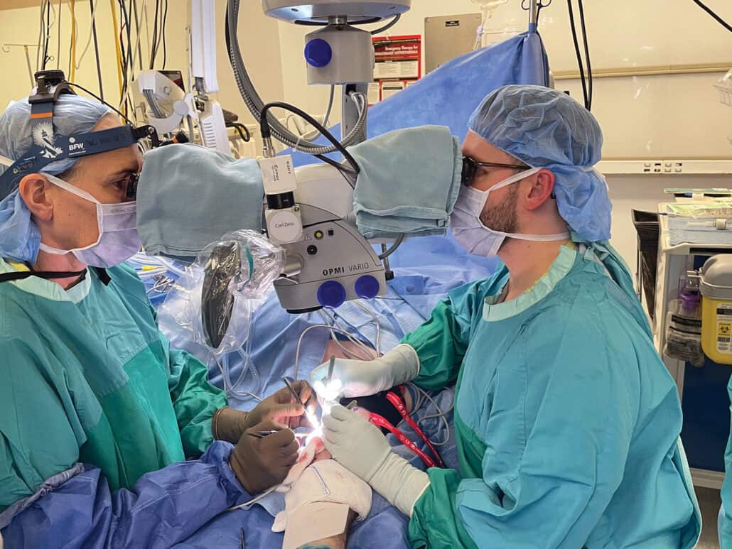
Photograph courtesy of Dr. Claire Temple-Oberle.
Lymph nodes act like small filtering stations located along the body’s lymphatic system. The lymphatic system is a network of vessels that drain excess fluid from tissues, removing harmful substances before returning clean fluid to the bloodstream. When surgeons remove lymph nodes, often from the armpit or groin, to check for or remove cancer, the limb near the surgical site can swell permanently. This is a common condition known as lymphedema.
In breast cancer patients, roughly 30 per cent who have their lymph nodes removed along with radiation develop it. Lymphedema brings swelling, heaviness and pain, and can mean a lifetime of compression bandages, lymphatic massages and physiotherapy, along with a higher risk of infection.
Dr. Claire Temple-Oberle, a University of Calgary professor in the departments of oncology and surgery, is focused on stopping lymphedema before it starts. As both a plastic surgeon and a surgical oncologist specializing in melanoma, she brings a dual vantage point to the problem, performing a preventative microsurgical technique when lymph nodes are removed.
“The number one way that we try to prevent lymphedema is to [avoid subjecting] patients to lymph node dissection operations,” she says. But some patients still require one, including women with inflammatory breast cancer and patients with bulky nodes from skin cancers like melanoma.
For those patients, Temple-Oberle intervenes during the dissection itself, performing a microsurgical procedure called immediate lymphatic reconstruction. As surgical oncologists remove the nodes, she protects the tiny lymphatic channels left behind, stretching them and preserving nearby veins from being cut too short. Then, under a microscope, she joins the lymphatic channels directly to those veins, a technique called lymphaticovenous anastomosis, which gives the disrupted lymphatic system a new drainage route.
Promising results
“We didn’t invent the procedure, but it wasn’t very common,” Temple-Oberle says, adding her team has performed the procedure since 2020. Now her Calgary team is leading research to prove it works. A randomized controlled trial for melanoma patients enrolled 40 participants, with final results expected next year. The technique has also been offered, through separate trials, to patients at the Arthur J.E. Child Comprehensive Cancer Centre needing lymph node dissections, including patients with breast, skin, rectal and ovarian cancers.
“The results have been really quite incredible,” Temple-Oberle says. The early numbers show a two-thirds reduction in lymphedema across the board. In breast cancer patients, the expected 30 per cent risk drops to roughly 10 per cent, and swelling that does occur tends to be mild rather than severe.
Temple-Oberle credits the team behind the work. Together, surgical oncologists, plastic surgeons, and operating room and clinic nurses collaborate on a procedure that adds time and complexity to surgeries. “Everyone really believed in this procedure. They jumped in enthusiastically,” she says.
As cancer survival rates climb, quality of life after treatment matters. Patients cured of cancer, Temple-Oberle says, shouldn’t have to live with the lasting symptoms of lymphedema, especially when a solution can be built into the first operation.
Built from scratch
A made-in-Alberta cancer drug’s journey to reach patients.

While Alberta is a leader in cancer clinical trials, most cancer drugs used here were discovered somewhere else, developed for years in labs far from the patients who eventually receive them. CCI-001 is different. This novel chemotherapy drug exists because Dr. Jack Tuszynski, the Allard Chair in Experimental Oncology at the University of Alberta’s Cross Cancer Institute (Cross) from 2005 to 2020, decided to build a cancer drug from the ground up, starting on a computer.
Underlying the drug’s discovery is a basic question: what makes cancer cells divide endlessly when healthy cells don’t? The answer involves microtubules, the rod-like structures that pull chromosomes apart during cell division, and a specific building block of them, beta III tubulin, that’s overexpressed in almost all cancer cells, but largely absent elsewhere in the body.
Existing tubulin-targeting drugs, like the widely used chemotherapy paclitaxel, didn’t distinguish well enough between cancerous and healthy cells. So, in 2010, Tuszynski’s team turned to an unlikely starting point: colchicine, a compound known to ancient Egyptians and still used for gout, but never successful as a cancer drug because of its toxicity.
Using computer modelling, his team redesigned the molecule, tuning its structure to bind tightly to the cancer-associated form of the protein, while leaving the healthy version alone.
It took roughly three years of computational work — using about 1,000 computers, Tuszynski says — before a physical compound was synthesized. A patent battle followed before CCI-001 was protected in 2016.
A Dutch lab tested the drug on different cancer cells, and found it worked best as a form of treatment for metastatic bladder cancer and pancreatic cancer. In mouse models of bladder cancer, CCI-001 outperformed the standard of care astoundingly well.
“We’re not shouting from the rooftops yet, but we are very happy that the results are supportive of our hypothesis,” Tuszynski says.
From bench to bedside
The multi-year project required sustained funding, including from the Alberta Cancer Foundation and through the Allard Foundation, sponsors of the Allard Chair. And when it entered human clinical trials in Edmonton in 2021, it marked a milestone: the first made-in-Alberta drug that kills cancer cells directly to be used in a clinical trial.
Medical oncologists Dr. Michael Sawyer and Dr. Jennifer Spratlin enrolled patients for the 2021 trial. Spratlin says watching Tuszynski’s drug move from computer model to hospital treatment room has been a career highlight. “
This has, in my entire career, been the coolest experience, to see something that Dr. Tuszynski did and bring it to Albertans,” she says, calling it “a wonderful story of bench to bedside.”
The trial is still technically in Phase 1, Sawyer says, though it has shifted from testing CCI-001 on its own to testing it in combination with standard chemotherapies. Results have been strong enough that the team hopes to eventually move the drug into a Phase 2 or Phase 3 trial. Unlike paclitaxel, CCI-001 isn’t causing the nerve damage that often forces patients off standard chemotherapy.
Tuszynski’s team has already made 1.4 kilograms of the compound, enough to treat 1,000 patients or more, in anticipation of a larger trial. So far in the human trial, researchers have seen responses primarily in ovarian, head and neck, lung and endometrial cancers. The trial also includes pancreatic and bile duct cancers, which researchers believe could respond to CCI-001.
Tuszynski sees the milestone as more than just one drug’s success. It’s proof Alberta can build a cancer drug from scratch and evidence of how powerful computational methods can be in finding new ones. After 15 years dedicated to CCI-001, he says his first emotion is gratitude, to the many people who helped along the way, and to the Allard family in particular for their funding.
But he’s not calling it finished. Tuszynski and others are now applying the same computational approach to search for new drugs for metastatic prostate cancer. The model behind CCI-001 isn’t a one-off, but a template for what comes next.